Abstract
The results of a theoretical study of the role of biophotons in electromagnetic process of realizing the phenomenon of life are presented in the article. This article is a continuation of the theoretical study of the team of authors, which was published in the Journal of Complexity in Health Sciences, Vol. 5, Issue 1, 2022, p. 22-34 and Vol. 5, Issue 2, 2022, p. 45-57. The aim of the theoretical study was to generalize the available scientific physical and biological knowledge of modern science about the role of biophotons in the electromagnetic processes of the phenomenon of life at the cellular level in order to deepen the fundamental knowledge of Complex Medicine. This study is a fragment of research work on “Development of algorithms and technologies for implementing a Healthy Lifestyle in patients with Noncommunicable Diseases based on the study of functional status” (state registration number 0121U108237: UDC 613 616-056-06: 616.1/9-03). General scientific methods and theoretical methods were used in this theoretical study. Based on the results of this fragment of the theoretical study, twelve conclusions were formulated. The formulated conclusions conceptualize such basic questions as: all living cells at the nanoscale consist of electromagnetic fields and generate electromagnetic fields, cell signaling and all cellular processes are due to electromagnetic interactions, ultra-weak photon emission is a universal optical phenomenon for all cells and plays an important role in communication and in cell life throughout the body.
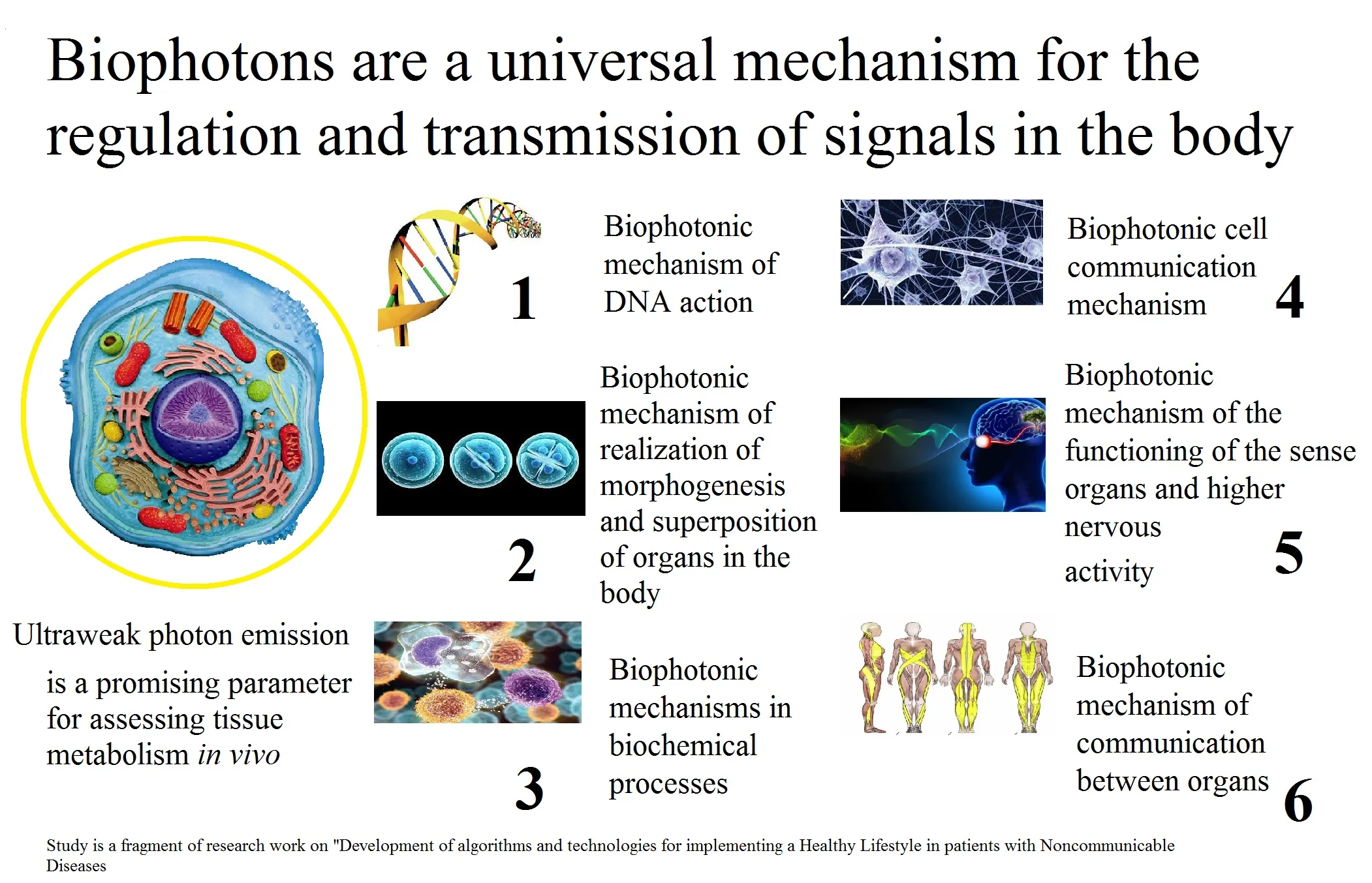
Highlights
- Biophotons are a universal mechanism for the regulation and transmission of signals in the body
- Ultraweak photon emission (yellow circle)
- Key mechanisms of biophotons (right)
Daring ideas are like chessmen moved forward. They may be beaten, but they may start a winning game
Johann Wolfgang von Goethe
1. Introduction
The question of what mechanisms and processes make biological molecules, tissues and organs alive is one of the most important for systems biology and complex medicine. Undoubtedly, the electrical activity of membranes and the quantum properties of water [1]-[4] play an important role, but this does not give a complete answer to this question [5]. The aspect of cellular communication is also a topical issue for understanding the phenomenon of biological life by science as well. Until recently, it was believed that cells interact with each other only due to the regulatory role of the nervous system and chemical interactions. However, it turned out that this is not the case and there are other mechanisms of intercellular interaction and cell signaling - electromagnetic processes of energy and information transfer [5]-[13]. Now science has accumulated a significant amount of scientific data describing these processes [10], [14-18]. It is necessary to integrate modern concepts of biophysical ideas about the structure and functioning of living cells of the human body into orthodox medical knowledge, namely, that all cells at the micro level of their structure consist of electromagnetic fields. It is categorically important to understand that all chemical reactions occurring in the cell have an electromagnetic quantum nature of the basis of the interaction process. The process of functional and morphological association of cells into tissues in living organisms is also due to electromagnetic interactions between cells. Significant scientific material has now been accumulated, confirming the presence and regulatory significance of electromagnetic fields in living biological organisms, including humans. Understanding by scientists the essence of the processes of quantum interaction between cells at the nanolevel of cellular organization in a healthy and diseased organism will improve the understanding of the pathogenesis of diseases, will allow finding new methods for their treatment and prevention.
The fundamental particle of the electromagnetic field and its quantum is the photon. A photon has the properties of an electromagnetic wave and a massless particle. Therefore, a photon can move at the speed of light and carry information [15]-[17], [19]-[20]. After the discovery and recognition of the fact that the cells of all living organisms and humans are constantly luminous, that is, they emit photons, the concept of “biophoton” was introduced. This phenomenon is called “ultra-weak photon emission” (UPE) [12], [21]-[23]. The discovery and study of UPE has become a new era in fundamental science and has given medicine a new potential for its further development.
Therefore, the aim of the theoretical study was to generalize the available scientific physical and biological knowledge of modern science about the role of biophotons in the electromagnetic processes of the phenomenon of life at the cellular level in order to deepen the fundamental knowledge of Complex Medicine.
2. Materials and methods
The analysis of the presented data is a fragment of research work of the Department of Internal Medicine and Emergency Medicine of Poltava State Medical University (23, Shevchenko St., 36011, Poltava, Ukraine) on “Development of algorithms and technologies for implementing a Healthy Lifestyle in patients with Noncommunicable Diseases based on the study of functional status” (state registration number 0121U108237: UDC 613 616-056-06: 616.1 / 9-03).
Scientific work is carried out in conjunction with the following scientific institutions: 1) Lithuanian University of Health Sciences (9, A. Mickevičius St., LT-44307, Kaunas, Lithuania), the cooperation coordinator is Head of Nephrology Department, prof., DM I.A. Bumblyte; 2) Shupyk National Healthcare University of Ukraine (9, Dorogozhytska St., 04112, Kiev, Ukraine), the cooperation coordinator is the Head of the Department of Informatics, Information Technologies and Transdisciplinary Education, prof., DM O.P. Mintser.
General scientific methods (dismemberment and integration of elements of the studied system, imaginary experiment, logical, historical research, analysis, induction, deduction and synthesis of knowledge) and theoretical methods (method of constructing theory, logical methods and rules of normative nature) were used in this theoretical study.
3. Results and discussion
When performing a theoretical study, it was found that the development of the idea of electromagnetic signaling lasted about 100 years: hypotheses and the first theories were formulated at the beginning of the twentieth century [24]-[26]. In 1924, for the first time, the fact of the presence of a distant electromagnetic interaction between cell cultures was experimentally established [27]. However, such revolutionary ideas could not at that stage be accepted by the paradigm of science of that time. It took 100 years for scientific truth to triumph in the 21st century thanks to a significant breakthrough in the fundamental sciences and the globalization of scientific knowledge. Now a large amount of results of scientific research makes it possible to make logical systemic generalizations and create on this basis a unified theory of metabolism [3], [4].
When analyzing and systematizing modern scientific knowledge, it was found that the cells of all living biological organisms at the micro level of their structure consist of electromagnetic fields [19], [28]-[29]. The vital activity of cells, including the processes of deterministic self-organization of molecules, functional and morphological association of cells in tissues, cellular regeneration, cellular activity, cellular metabolism, occurs due to quantum information electromagnetic interaction [electromagnetic signaling] due to the transport of energy in various forms and packages (electrons, photons, solitons, etc.) [3]-[5], [7]-[10], [14], [16], [19], [30]-[33]. And this ensures the phenomenon of their collective biological life in the body. It was found that biophotonic mechanisms are the most important manifestation of electromagnetic signaling. [6, 12]. In the body, it is electromagnetic energy [photons] that is the necessary target final substrate of biochemical metabolism, which provides all the basic processes of cell vital activity, including the functioning of the deoxyribonucleic acid (DNA) molecule with the formation of an electromagnetic coherent cellular biological state [5], [11], [34]-[36].
The unique physiological role of biophotons in the life processes of living organisms and humans is due to the quantum mechanical features of the functioning of living cells. To explain and to further integrate these ideas into integrated medicine, we have conceptualized an adapted description of a simplified view of this. It is important to understand that the process of functional and morphological integration of cells into tissues in living organisms, all chemical reactions occurring in cells and the phenomenon of their biological life are due to electromagnetic interactions. This is so because if we “immerse ourselves in the Nano world” and reduce the scale to 10-9-10-13 cm, then we will not see the cell and matter in the sense familiar to the human imagination. After reducing the scale to the level of the Nano world, there are no atoms, but only the energy of the movement of electromagnetic waves of the internal environment. On the indicated scale, the atomic nucleus is an electromagnetic process, which is conditionally described as a rotating electromagnetic rod, consisting of protons and neutrons. Protons and neutrons are magnetic waves moving at the speed of light along a spiral ring path. Further reduction of the scale to 10-28-10-35 cm corresponds to the description of bosons in the form of rings of wave processes linked by magnetic and other interactions into ordered structures, with a speed of movement 106 greater than the speed of light, etc. Thus, modern science has discovered that all atoms and molecules are an organized form of electromagnetic energy and all interactions between atoms and molecules in cells occur due to electromagnetic energy as well. It was also discovered that cells generate biological electromagnetic fields in the ultraviolet and in the visible range of the spectrum, as well as in the frequency range below the terahertz range [3]-[5], [17], [20], [37]-[43]. If the frequency of the oscillatory charge is high and approaches the optical part of the spectrum of the electromagnetic field, then the electromagnetic waves generated by cells [or cell organelles] begin to show their corpuscular properties when interacting with matter - then we can talk about the generation of light particles or biophotons, which are a scientifically established physical substrate cellular electromagnetic signaling [34], [44], [45].
In 1976, the term “biophoton” (from the Greek "βίος" – life and "Φως" – light, power) was introduced into scientific use by the German biophysicist F.-A. Popp [21], [46]. Scientific research led F.-A. Popp to the understanding that light is produced inside a biological organism, although this did not fit into the then scientific dogma. F.-A. Popp, as the founder of the International Institute of Biophysics (Düsseldorf, Germany), organized an international research network of 19 research institutes in 13 countries, which was engaged in biophotonic research. The contribution of F.-A. Popp to fundamental science lies in the fact that by his research he formed an adequate level of scientific evidence for the existence of the phenomenon of “weak photon emission” in all living cells and created the necessary theoretical basis for further research in this direction [46]-[51].
As a result of our theoretical study, we have formulated such concepts for complex medicine:
– biophoton emission or Ultra-weak Photon Emission (UPE) is a scientifically proven optical phenomenon of electromagnetic radiation in the spectral region from 200 to 800 nm, which really exists and is universal for most living biological systems, at a constant rate from several photons per cell per day to several hundred photons per organism per day [22], [23], [44], [47], [52]-[58];
UPE is invisible to the human eye [threshold of human vision ~ 1·Е6 s-1 cm-2], has low intensity, higher energy compared to thermal or conventional chemical activation [the range of energy values is 1.67-3.41 eV; in the spectral range < 700 nm UPE is 1·E10 higher than the statistical Boltzmann distribution, has a value of ~10-1·E4 photons per s-1·cm-2 and its sources are non-statistical, thermal equilibrium phenomena] [22], [23], [44], [47]-[51], [56]-[62];
– UPE is involved in the implementation of the process of cell life and is of key importance in this case, it correlates with indicators of metabolism, hormonal levels, with the chronobiological rhythms of living organisms, and the activity of electromagnetic fields of the near space [22], [23], [40], [46]-[48], [52], [54], [55], [57], [61], [62]-[65];
– UPE is the result of the space-time manifestation of the energy of the biological electromagnetic field of a living cell, the coherent properties of which are electromagnetic intercellular signaling [22], [23], [35], [45], [47], [52], [57], [59], [62].
According to biophysicists, light and living matter are closely related. Indeed, light and living matter have such a special relationship that is now being advanced to the most advanced frontiers of modern research in the field of quantum computing, optics and other nonlinear optical phenomena in condensed matter physics [14].
Biophotons are connected with the energy-matter interaction: the absorbed electromagnetic radiation leads to the excitation of the state of the atom (quantum jumps) and vice versa. To establish a transmolecular bond, the molecules involved must be in some kind of excited state [14]. Classical observations in solid-state systems suggest that relaxation can occur in certain quantum steps, i.e. an electron can go to a lower but still excited state without emitting visible light, or can start to move, thus becoming an electric current, and in in other cases, it may be involved in a chemical reaction. Complete relaxation to the ground state is recombination with the remaining positively charged hole behind and this will result in the emission of electromagnetic energy. It is known from quantum electrodynamics that orbiting electrons and the nucleus constantly exchange virtual (not quite real) photons. However, in biotic systems, an excited electron-hole pair, or exciton, can travel long distances within the system before releasing energy by emitting a photon. It is believed that the formation of excitons and their distribution is involved in the main energy conversions and in biocommunications. In fact, the DNA molecule itself is considered to be an excited duplex or exciplex system in which photons are effectively stored between two DNA strands. Therefore, living systems emit light from processes occurring in all cells [40], [46], [52], [56], [57].
The correctness of this is confirmed by such an interesting fact for complex medicine that in situ at least 75 % of biophoton activity comes from cell DNA.
This phenomenon was tried to be studied, but biophoton DNA was inactive during isolation and purification. This was found such a scientific explanation: in DNA, photons are in a state of Bose-Einstein condensate. DNA has an information density that is 1·109 higher than any known technical solution to date [66], [78]. This high density of information leads to a phenomenon known in physics as a Bose-Einstein condensate: in this phenomenon, photons are captured in a “cryotrap”, compacted and “frozen” in time. The stored light constitutes the elemental stability of the DNA molecule. It is assumed that 97.98 % of inactive human DNA together with “frozen” energy plays an essential role in the organization of 2.02 % of genetically expressed DNA. Consequently, an electromagnetic coherent cellular biological state is established in the form of a Bose-Einstein condensate, in which photons of the same frequency and phase are aligned with each other. Thus, the range of interaction is increased from microscopic to including macroscopic entities such as cells, organs, whole organisms, and even more [52], [66]. This confirmed H. Fröhlich’s assumption about the biophotonic mechanism of DNA action, which he made back in the 50s of the twentieth century [41], [46], [47], [50].
The second fundamental biological role of biophotons is the implementation of the processes of morphogenesis. UPE is involved in the formation of the morphogenetic field. This is a chemical-mechanical-electromagnetic process that has a holistic effect on DNA, and this controls the growth, differentiation and coordination of molecules in cells. The morphogenetic field can deform larger molecules by changing electric fields, changing chemical potentials, and controlling molecular behavior. The weakening of the morphogenetic field causes chaotic morphogenesis. This was confirmed in experiments carried out with Drosophila embryos exposed to weak electric and magnetic fields, which weakened the morphogenetic field, which caused malformations [14], [66]. A detailed observation of mitosis showed that there is a correspondence between the structural pattern of the mitotic apparatus and the lines of the electric field. At any moment of this process, a spatial distribution of energy is detected inside the cell, which controls the flow of chemical reactions in a well-coordinated functional sequence. As a result, it has been proposed to compare mitosis with a technical resonator. Evidence has been obtained that mitotic patterns are excellent examples of long-lived photonic storage units in biological systems [12], [52], [65], [66].
To date, a mechanism for the participation of UPE in the implementation of genetic information has been developed and there is a model for regulating life through UPE in living organisms [8], [9], [47], [49], [65]. It can be simplified as follows: in the nucleus there is a spiral-shaped genetic material that functions like a biological laser, receiving energy in the cell from nutrients in the form of photons [49], [67]. The same studies indicate that normal human cells have the ability to accumulate the ultra-weak light energy transmitted to them and use it for their complex biochemical processes. For example, dying cells lost this storage capacity and showed a significant increase in UPE before death. This is called the “death flash” [68]. It has now been established that a similar state is gradually formed in the cells in the process of aging of the organism. Moreover, in the case of cancer cells, cellular toxins accumulate over many years and often lead to the deposition of cellular debris in the tissues (for example, vascular atherosclerosis), which also disrupts the accumulation and causes changes in UPE [69], [70].
Contrary to the common assumption that molecular reactivity is determined by the chaotic stimulation of thermal energy, it is now becoming clear that UPE is the result of the spatiotemporal manifestation of electromagnetic field energy.
Due to the coherence property of this biophoton field, cells manifest the ability to use this energy as a communication tool, without which a single cell, both in simple and complex multicellular organisms, could not communicate with others. In this sense, UPE coherence also includes communication processes that involve the whole organism. Because living systems are mutually related entities embedded in their respective environments, this process of conjugation results in a coherent interaction with all the cells that make up an organism. Thus, biophotons are a key tool in the processes of intercellular and transcellular communication [12], [52], [54].
The purpose and function of biophotons are also associated with the superposition of various cells inside the organ, since they form an information field that is transmitted through the connective tissue according to the fiber optic principle. Cell membranes are located in the nodal planes of the interference pattern. As can be seen from the cell cycle, the energy distribution of the extracellular space serves as a means of communication and interaction in regulatory processes with neighboring cell units. In this regard, the connective tissue with its network of collagen fibers plays a fundamental role in the transport of electromagnetic energy. Traditional classical concepts of connective tissue assign it only a binding role that connects tissues together and maintains the flexibility of a body part. However, connective tissue has a more fundamental role to play in biophotonics, as it is a “fiber optic” network that transmits optical messages throughout the body [14], [66]. The main generators of UPE are myocytes. The functioning of muscle cells is another example of how biophotonics is built into the whole human body. The molecules of myosin and actin are packed and arranged very precisely, approaching the regularity of piezocrystals. This creates the conditions for significant generation of biophotons by the muscles. The moment of muscle contraction involves electron tunneling (passing under an energy barrier that occurs within a nanosecond) with coordinated fluctuations. Muscle cell contraction occurs in definite and synchronous quantum steps, and this is a fluctuation-free chain process of sliding of actin-myosin filaments – this is essentially a characteristic of a coherent quantum field. At the same time, a very large number of cells involved in a typical muscle contraction perform the same milling of molecular filaments in a consistent manner on a scale of distances spanning nine orders of magnitude. In addition, the contraction of the muscles of the body occurs almost constantly, which converts energy to the level of biophotons. At the same time, electromagnetic energy in the muscles can be produced in the amount necessary for the body with almost 100 % efficiency [14], [66]. The important point is that the muscles produce biophotons for the entire human body. The connective tissue framework is in the muscle fibers, which in the body form a single myofascial system, and therefore biophotons can be redistributed throughout the body to the “fiber optic channel” of the connective tissue. This connective tissue system is called the “primary vascular system”. Its main function is considered to be the transport of biophotons [71]-[75]. This is another new promising direction for research in complex medicine.
Biophotons are a regulatory mechanism common to all processes due to the fact that they are involved in signal transmission and are used to amplify very weak stimuli. The nervous system of the retina, for example, has time constants of the order of 10·102 s – this is too slow, given the actual speed of visual perception, to activate a single phosphodiesterase molecule after absorbing a photon. Most of the amplification is actually at the initial stage, when single-photon excited rhodopsin passes to excitation of at least 500 transducin molecules within 1 ms. While the underlying mechanism is still the subject of current research, it is hypothesized that biophotonic processes are involved [14], [52], [55], [66].
All of the above indicates the participation of UPE in the processes of cellular regeneration, cellular activity, cellular metabolism, viability and replication [45], [55], [65], [70], [76], [77]. Biophotonic activity of cells controls chemical reactions in the cytoplasm, provides resonant signaling between cells and cell regeneration processes. As in solid-state systems, the superposition of different modes in the optical range of electromagnetic radiation gives an accurate spatial resolution of the picture of the intensity of “standing waves” - solitons. A spatially distributed electric field guides molecules and accurately drives over 100·103 chemical reactions per second [12]. The cytoplasm provides only a part of the biophoton activity due to the activity of microtubules involved in the multiplication of biophoton radiation coming from the cell nucleus. Microtubules, along with contact communication junctions, conduct biophoton impulses to neighboring cells and to the extracellular matrix. Adhesive forces between cells connect them to functional blocks and thus make it possible to form a resonator system also for long-wavelength photons. When a cell of such a unit dies, the resonant frequency is disturbed and some photons are emitted, thereby initiating the process of cellular regeneration [52], [66], [78]. The existence of a relationship between UPE levels and reactive oxygen species in the tissues of living biological systems has been proven [66], [69]. UPE are involved in the photochemical processes of the human eye [79], [80]. There are theoretical prerequisites to believe that it is UPE that allows you to create internal biophysical pictures with visual perception and imagination in humans [22], [23], [47], [50], [66], [81], [82].
It has been established that healthy tissues and organisms have stronger and more stable UPE radiation than damaged and diseased ones. This became the basis for studying the role of UPE as another functional parameter for assessing the vital activity of a living biological system, including humans [36], [60], [62], [83]-[89].
Biophoton emission and UPE studied in many tissues [24], [60], [61], [70], [76], [90]-[93] and in some human diseases [55], [64], [80], [83], [84], [87], [94]-[99].
Our own studies in patients with chronic non-communicable diseases using the electrophoton emission analysis established significant differences (0.0001) in the intensity and area of luminescence parameters between groups of patients and healthy respondents [97]-[98], [100]-[101]. This once again confirms the fact that metabolic electromagnetic processes in the human body during pathology change significantly at the quantum level and methods for clinical assessment of biophoton levels can be used as a promising new clinical indicator.
Studies carried out on representatives of the plant, fungal, and animal kingdoms have also confirmed the fact that UPE changes in normal and pathological conditions in living organisms. Thus, in a significant number of studies, it was found that in plants, healthy leaves radiated UPE much more strongly and more slowly at any given time than wilted and diseased leaves [22], [62], [102].
It was found that UPE values changed in protozoa, plants, lichens and fungi after stress (chemical) [22], [62], [66]. Similar results were obtained in studies of representatives of the animal kingdom [81], [82], [103]-[107].
There is a relationship between UPE parameters and exposure of living biological systems to natural sunlight and active movement. The positive energizing effect of natural sunlight under the conditions of living organisms in the natural environment has been established [66], [108].
The UPE study of organic food showed that this parameter reflects their quality/freshness and biological differences of food as a conditionally living system. The phenomenon of freshness of the product is due to the presence in it of the basic electromagnetic processes of the phenomenon of biological life - electromagnetic fields of cells that have not yet undergone chemical decay and decomposition by microorganisms/decay, maintain integrity, organoleptic properties [51], [109]-[112]. By investigating UPE, science now has the opportunity to judge the biological quality of food products from the position of basic electromagnetic processes associated with the very essence of the phenomenon of life of biological molecules. In the future, UPE may become a new objective criterion for the quality and biological freshness of food products, based on the flow of energy processes at their micro level.
Biophotons and UPE as an existing phenomenon deepen the scientific understanding of the phenomenon of biological life and their role in this phenomenon of life, being not only a product and result of metabolic processes at the micro level of metabolism, but also performing an energizing and regulatory role as a component of the electromagnetic signaling mechanism.
4. Conclusions
The following conclusions can be drawn on the basis of the results of the theoretical study:
1) All cells and all cell-forming organelles are composed of electromagnetic fields at the micro level of their structure.
2) In cells, the processes of deterministic self-organization of molecules are realized due to quantum informational electromagnetic interaction due to the transport of energy in various forms and packages (electrons, photons, solitons, etc.), which ensures the phenomenon of its biological life.
3) The process of functional and morphological association of cells into tissues in living organisms, all chemical reactions occurring in cells and the phenomenon of their biological life are due to electromagnetic interactions.
4) Cells generate biological electromagnetic fields in the ultraviolet and visible range of the spectrum, as well as in the frequency range below the terahertz range. If the frequency of the oscillatory charge is high and approaches the optical part of the spectrum of the electromagnetic field, then the electromagnetic waves generated by cells [or cell organelles] begin to show their corpuscular properties when interacting with matter - then we can talk about particles of light or biophotons, which are a scientifically established physical substrate of cellular electromagnetic signaling.
5) Emission of biophotons or UPE is a universal optical phenomenon for most living biological systems, including humans, consisting of electromagnetic radiation in the spectral region from 200 to 800 nm, with a constant rate from several photons per cell per day to several hundred photons per organism per day. UPE accompanies the process of life, is of key importance for the life of cells, correlates with indicators of metabolism, hormonal levels and chrono biological rhythms of living organisms. UPE is the result of the spatio-temporal manifestation of the energy of the biological electromagnetic field of a living cell, the coherent properties of which are electromagnetic intercellular signaling.
6) In situ, at least 75 % of biophoton activity comes from DNA. In the nucleus is a spiral-shaped genetic material that functions like a biological laser, receiving energy in the cell from nutrients in the form of photons.
Photons stored in DNA in the form of a Bose-Einstein condensate constitute the elementary stability of the DNA molecule, establish an electromagnetic coherent cellular biological state in which photons of the same frequency and phase are aligned with each other. This allows us to assert that those 98 % of DNA that were considered "junk" are responsible for the implementation of complex electromagnetic processes inside the cell through the biophoton mechanism.
7) Normally, human cells are characterized by the ability to accumulate the ultra-weak light energy transmitted to them [biophotons] and use it for biochemical processes. The ability to accumulate photon energy decreases under pathological conditions and aging, which is objectively manifested by a change in the processes of emission of biophotons during registration.
8) Biophoton emission or UPE provides resonant signaling between cells, processes of cellular regeneration, cellular activity, cellular metabolism, viability and replication.
9) The emission of biophotons or UPE [cell signaling and cellular biochemical processes, respectively] strongly correlates with the cell cycle and other functional states of cells and organisms, and manifests itself as a response to many external stress stimuli, depends on the influence of external natural electromagnetic fields (chrono biological rhythms, geomagnetic influences, influences of near space).
10) The emission of biophotons or UPE can play an important potential role in specialized cells of the nervous system in the transmission and processing of nerve signals, being one of the mechanisms of the higher functions of the nervous system of complex living biological organisms, including humans, and presumably can make it possible to create internal biophysical pictures during visual perception and imagination in a human.
11) The emission of biophotons or UPEs are involved in tissue morphogenesis, providing a superposition of various cells inside the organ, since they form an information field that is transmitted through the connective tissue according to the fiber optic principle.
12) The emission of biophotons or UPE is a manifestation of the final link of metabolism and is an energy and information carrier that is transmitted through the connective tissue of the whole organism according to the fiber optic principle.
This fundamentally deepens the scientific understanding of the mechanisms of the cellular level of organization and functioning of the human body, forms a new view on the description of biological processes that occur in the human body in normal and pathological conditions, expands the possibilities of in-depth study, description of the pathogenesis of human diseases and demonstrates the feasibility of a paradigm transition from electrochemical concept of metabolism to magnetoelectrochemical. Estimation of photon emission parameters can be of great practical importance for use in biomedical research and in the food industry to determine the quality of a biological substrate [if we are talking about food, etc.].
References
-
G. Nevoit, I. A. Bumblyte, M. Potyazhenko, and O. Minser, “Modern biophysical view of electromagnetic processes of the phenomenon of life of living biological systems as a promising basis for the development of complex medicine: the role of cell membranes,” Journal of Complexity in Health Sciences, Vol. 5, No. 1, pp. 22–34, Jun. 2022, https://doi.org/10.21595/chs.2022.22787
-
G. Nevoit, I. A. Bumblyte, M. Potyazhenko, and O. Minser, “Modern biophysical view of electromagnetic processes of the phenomenon of life of living biological systems as a promising basis for the development of complex medicine: the role of water,” Journal of Complexity in Health Sciences, Vol. 5, No. 2, pp. 45–57, Dec. 2022, https://doi.org/10.21595/chs.2022.23089
-
O. P. Minser, M. M. Potyazhenko, and G. V. Nevoit, Magnetoelectrochemical Theory of Metabolism. (in Ukraine), Kyiv-Poltava: Interservice, 2021.
-
O. P. Mintser, M. Potiazhenko, A. L. Vainoras, I. B. Bumblytė, and G. V. Nevoit, “Informational Analytical Representations of the Magneto-Electrochemical Theory of Metabolism, Life and Health,” Ukraïnsʹkij Žurnal Medicini, Bìologìï Ta Sportu, Vol. 7, No. 5, pp. 232–246, Nov. 2022, https://doi.org/10.26693/jmbs07.05.232
-
E. Schrödinger, What is Life? The Physical Aspect of the Living Cell. Cambridge: University Press, 1944.
-
M. Cifra, J. Z. Fields, and A. Farhadi, “Electromagnetic cellular interactions,” Progress in Biophysics and Molecular Biology, Vol. 105, No. 3, pp. 223–246, May 2011, https://doi.org/10.1016/j.pbiomolbio.2010.07.003
-
A. S. Davydov, “Solitons as energy carries in biological systems,” Studia Biophys, Vol. 62, No. 1, pp. 1–8, 1977.
-
H. Frohlich, “Long-ranch coherence and energy storage in biological systems,” International Journal of Quantum Chemistry, Vol. 2, No. 3, pp. 641–649, 1968.
-
H. Frohlich and F. Kremer, Coherent Excitations in Biological Systems. Berlin: Springer-Verlag, 1985.
-
G. R. Fleming, G. D. Scholes, and Y.-C. Cheng, “Quantum effects in biology,” Procedia Chemistry, Vol. 3, No. 1, pp. 38–57, 2011, https://doi.org/10.1016/j.proche.2011.08.011
-
Felix Scholkmann, Daniel Fels, and Michal Cifra, “Non-chemical and non-contact cell-to-cell communication: a short review,” American Journal of Translational Research, Vol. 5, No. 6, p. 586, 2013.
-
R. Vanwijk, “Bio-photons and Bio-communication,” Journal of Scientific Exploration, Vol. 15, No. 2, pp. 183–197, 2001.
-
K. Yamanouchi, Quantum Mechanics of Molecular Structures. Berlin Heidelberg: Springer-Verlag, 2016.
-
M. W. Ho, The Rainbow and the Worm: The Physics of Organisms. World Scientific Publishing Company, 2003.
-
K. M. Merz, “Using quantum mechanical approaches to study biological systems,” Accounts of Chemical Research, Vol. 47, No. 9, pp. 2804–2811, Sep. 2014, https://doi.org/10.1021/ar5001023
-
J. Mehra, “Quantum mechanics and the explanation of life: the inclusion of human consciousness in quantum physics recognizes mind as the primary reality: Will a new science arise that can harmonize quantum physics and biology?,” American Scientist, Vol. 61, No. 6, pp. 722–728, 2021.
-
K. A. Peacock, The Quantum Revolution. Greenwood: Publishing Group, 2008.
-
H. A. Pohl and J. K. Pollock, “Biological Dielectrophoresis,” Modern Bioelectrochemistry, pp. 329–376, 1986, https://doi.org/10.1007/978-1-4613-2105-7_12
-
Encyclopedia of Physical Science and Technology Reference Work. Academic Press, 2001.
-
M. K. Gaillard, P. D. Grannis, and F. J. Sciulli, “The standard model of particle physics,” Reviews of Modern Physics, Vol. 71, No. 2, 1999.
-
F. A. Popp, Biophotonen e Ein neuer Weg zur Lösung des Krebsproblems. Verlag fúr Medizin Dr. Ewald Fischer, 1976.
-
F.-A. Popp, K. H. Li, and Q. Gu, Recent Advances in Biophoton Research and Its Applications. Singapore: World Scientific Publishing, 1992.
-
F. A. Popp, J. J. Chang, A. Herzog, Z. Yan, and Y. Yan, “Evidence of non-classical (squeezed) light in biological systems,” Physics Letters A, Vol. 293, No. 1-2, pp. 98–102, Jan. 2002, https://doi.org/10.1016/s0375-9601(01)00832-5
-
H. J. Niggli, “Biophotons: ultraweak light impulses regulate life processes in aging,” Journal of Gerontology and Geriatric Research, Vol. 3, No. 2, p. 143, 2014, https://doi.org/10.4172/2167-7182.1000143
-
H. S. Burr and F. S. C. Northrop, “The electro-dynamic theory of life,” The Quarterly Review of Biology, Vol. 10, No. 3, p. 322, 1935.
-
A. Gurwitsch, “Untersuchungen über den zeitlichen Faktor der Zellteilung,” Archiv für Entwicklungsmechanik der Organismen, Vol. 32, No. 3, pp. 447–471, Oct. 1911, https://doi.org/10.1007/bf02287040
-
A. Gurwitsch, “Physikalisches über mitogenetische Strahlen,” Archiv für Mikroskopische Anatomie und Entwicklungsmechanik, Vol. 103, No. 3-4, pp. 490–498, Oct. 1924, https://doi.org/10.1007/bf02107498
-
B. Carithers and P. Grannis, “Discovery of the top quark,” Beam Line, Vol. 25, No. 3, pp. 4–16, 1995.
-
B. C. Chauhan, М. Picariello, J. Pulido, and E. Torrente-Lujan, “Quark-lepton complementarity, neutrino and standard model data predict (θ13PMNS=9+1-2),” European Physical Journal, Vol. 50, No. 3, pp. 573–578, 2007.
-
E. Sjulstok, J. M. H. Olsen, and I. A. Solov’Yov, “Quantifying electron transfer reactions in biological systems: what interactions play the major role?,” Scientific Reports, Vol. 5, No. 1, pp. 1–11, Dec. 2015, https://doi.org/10.1038/srep18446
-
L. Demetrius, “Quantum statistics and allometric scaling of organisms,” Physica A: Statistical Mechanics and its Applications, Vol. 322, pp. 477–490, May 2003, https://doi.org/10.1016/s0378-4371(03)00013-x
-
V. P. Gupta, Principles and Applications of Quantum Chemistry. Elsevier, 2016, https://doi.org/10.1016/c2014-0-05143-x
-
P. Volpe, “Interactions of zero-frequency and oscillating magnetic fields with biostructures and biosystems,” Photochemical and Photobiological Sciences, Vol. 2, No. 6, pp. 637–648, Jun. 2003, https://doi.org/10.1039/b212636b
-
R. K. Adair, “Vibrational resonances in biological systems at microwave frequencies,” Biophysical Journal, Vol. 82, No. 3, pp. 1147–1152, Mar. 2002, https://doi.org/10.1016/s0006-3495(02)75473-8
-
Davies and P. C. W., “Does quantum mechanics play a non-trivial role in life?,” Biosystems, Vol. 78, No. 1-3, pp. 69–79, Dec. 2004, https://doi.org/10.1016/j.biosystems.2004.07.001
-
K. G. Korotkov, The Energy of Health. Amazon.com, 2019.
-
R. P. Bajpai, “Coherent nature of the radiation emitted in delayed luminescence of leaves,” Journal of Theoretical Biology, Vol. 198, No. 3, pp. 287–299, Jun. 1999, https://doi.org/10.1006/jtbi.1999.0899
-
R. P. Bajpai, “Quantum coherence of biophotons and living systems,” Indian Journal of Experimental Biology, Vol. 41, No. 5, pp. 514–527, May 2003.
-
R. P. Bajpai, “Biophoton emission in a squeezed state from a sample of parmelia tinctorum,” Physics Letters A, Vol. 322, No. 1-2, pp. 131–136, Feb. 2004, https://doi.org/10.1016/j.physleta.2003.12.050
-
M. Cifra, J. Pokorný, D. Havelka, and O. Kučera, “Electric field generated by axial longitudinal vibration modes of microtubule,” Biosystems, Vol. 100, No. 2, pp. 122–131, May 2010, https://doi.org/10.1016/j.biosystems.2010.02.007
-
G. Hyland, “Physics and biology of mobile telephony,” The Lancet, Vol. 356, No. 9244, pp. 1833–1836, Nov. 2000, https://doi.org/10.1016/s0140-6736(00)03243-8
-
H. A. Pohl, “Electrical aspects of cell growth and invasiveness,” Journal of Biological Physics, Vol. 7, No. 1, pp. 1–16, Jan. 1979, https://doi.org/10.1007/bf02349935
-
H. A. Pohl, “Electrical oscillation and contact inhibition of reproduction in cells,” Journal of Biological Physics, Vol. 9, No. 4, pp. 191–200, Dec. 1981, https://doi.org/10.1007/bf01988221
-
L. V. Beloussov, V. L. Voeikov, and V. S. Martynyuk, Biophotonics and Coherent Systems in Biology. Springer Science+Business Media, 2007.
-
L. V. Beloussov, “From molecular machines to macroscopic fields: an accent to characteristic times,” European Journal of Biophysics, Vol. 1, No. 1, 2013, https://doi.org/10.11648/j.ejb.20130101.12
-
J. J. Chang, J. Fisch, and F. A. Popp, Biophotons. Kluwer Academic Publishers, 1998.
-
F. A. Popp and L. V. Beloussov, Integrative Biophysics, Biophotons. Boston: Kluwer Academic Publishers, 2003.
-
R. van Wijk, E. P. A. van Wijk, J. Pang, M. Yang, Y. Yan, and J. Han, “Integrating ultra-weak photon emission analysis in mitochondrial research,” Frontiers in Physiology, Vol. 11, p. 540573, Jul. 2020, https://doi.org/10.3389/fphys.2020.00717
-
F. A. Popp, W. Nagl, K. H. Li, W. Scholz, O. Weingärtner, and R. Wolf, “Biophoton emission. New evidence for coherence and DNA as source,” Cell Biophysics, Vol. 6, No. 1, pp. 33–52, Mar. 1984, https://doi.org/10.1007/bf02788579
-
F. A. Popp, Coupling of Fröhlich-Modes as a Basis for Biological Regulation. University of Liverpool, 2006.
-
F. A. Popp, Die Botschaft der Nahrung. Frankfurt a.M. (FRG): Zweitausendeins, 2005.
-
M. Bischof, Biophotons, the Light in Our Cells. Frankfurt a.M. (FRG): Zweitausendeins, 1995.
-
M. Cifra and P. Pospíšil, “Ultra-weak photon emission from biological samples: Definition, mechanisms, properties, detection and applications,” Journal of Photochemistry and Photobiology B: Biology, Vol. 139, pp. 2–10, Oct. 2014, https://doi.org/10.1016/j.jphotobiol.2014.02.009
-
J. A. Ives et al., “Ultraweak photon emission as a non-invasive health assessment: a systematic review,” PLoS ONE, Vol. 9, No. 2, p. e87401, Feb. 2014, https://doi.org/10.1371/journal.pone.0087401
-
M. Kobayashi and H. Inaba, “Photon statistics and correlation analysis of ultraweak light originating from living organisms for extraction of biological information,” Applied Optics, Vol. 39, No. 1, pp. 183–192, Jan. 2000, https://doi.org/10.1364/ao.39.000183
-
M. Kobayashi, T. Iwasa, and M. Tada, “Polychromatic spectral pattern analysis of ultra-weak photon emissions from a human body,” Journal of Photochemistry and Photobiology B: Biology, Vol. 159, pp. 186–190, Jun. 2016, https://doi.org/10.1016/j.jphotobiol.2016.03.037
-
X. Shen and R. van Wijk, Biophotonics: Optical Science and Engineering for the 21st Century. Springer, 2005.
-
R. van Wijk and X. Shen, Biophotonics: Optical Science and Engineering for the 21st Century. Boston, MA: Springer US, 2005, https://doi.org/10.1007/b106475
-
M. Calcerrada and C. Garcia-Ruiz, “Human ultraweak photon emission: key analytical aspects, results and future trends – a review,” Critical Reviews in Analytical Chemistry, Vol. 49, No. 4, pp. 368–381, Jul. 2019, https://doi.org/10.1080/10408347.2018.1534199
-
M. Cifra, E. van Wijk, H. Koch, S. Bosman, and R. van Wijk, “Spontaneous ultra-weak photon emission from human hands is time dependent,” Radioengineering, Vol. 16, No. 2, p. 15, 2007.
-
R. van Wijk, M. Kobayashi, and E. P. A. van Wijk, “Anatomic characterization of human ultra-weak photon emission with a moveable photomultiplier and CCD imaging,” Journal of Photochemistry and Photobiology B: Biology, Vol. 83, No. 1, pp. 69–76, Apr. 2006, https://doi.org/10.1016/j.jphotobiol.2005.12.005
-
R. van Wijk, Light in Shaping Life-Biophotons in Biology and Medicine. Geldermalsen: Meluna Research, 2014.
-
A. V. P. Kaznacheev, L. P. Mikhailova, and N. B. Kartashov, “Distant intercellular electromagnetic interaction between two tissue cultures,” Bulletin of Experimental Biology and Medicine, Vol. 89, No. 3, pp. 345–348, Mar. 1980, https://doi.org/10.1007/bf00834249
-
M. Kobayashi, D. Kikuchi, and H. Okamura, “Imaging of ultraweak spontaneous photon emission from human body displaying diurnal rhythm,” PLoS ONE, Vol. 4, No. 7, p. e6256, Jul. 2009, https://doi.org/10.1371/journal.pone.0006256
-
W. Nagl and F. A. Popp, “A physical (electromagnetic) model of differentiation. 1. Basic considerations,” Cytobios, Vol. 37, No. 145, pp. 45–62, 1983.
-
P. Madl, “Biophotonics or the light of life. Lecture series: block I/IV – biophysics in life sciences. Of physics and biophysics,” University of Salzburg, 2006.
-
S. P. Sitko, “The realization of genome in the notions of physics of the alive", “Medical Data” Medical Review, Vol. 4, No. 2, pp. 207–216, 2012.
-
J. Slawinski, “Photon emission from perturbed and dying organisms: biomedical perspectives,” Complementary Medicine Research, Vol. 12, No. 2, pp. 90–95, 2005, https://doi.org/10.1159/000083971
-
E. van Wijk, M. Kobayashi, R. van Wijk, and J. van der Greef, “Imaging of ultra-weak photon emission in a rheumatoid arthritis mouse model,” PLoS ONE, Vol. 8, No. 12, p. e84579, Dec. 2013, https://doi.org/10.1371/journal.pone.0084579
-
Hugo J. Niggli, Salvatore Tudisco, Luca Lanzanò, Lee Ann Applegate, Agata Scordino, and Francesco Musumeci, “Laser-ultraviolet-A induced ultra weak photon emission in human skin cells: A biophotonic comparison between keratinocytes and fibroblasts,” Indian Journal of Experimental Biology, Vol. 46, No. 5, pp. 358–363, May 2008.
-
K. A. Kang, “Chronological review on scientific findings of bonghan system and primo vascular system,” Advances in Experimental Medicine and Biology, Vol. 923, pp. 301–309, 2016, https://doi.org/10.1007/978-3-319-38810-6_40
-
H. G. Kim, “Formative research on the primo vascular system and acceptance by the Korean scientific community: the gap between creative basic science and practical convergence technology,” Journal of Acupuncture and Meridian Studies, Vol. 6, No. 6, pp. 319–330, Dec. 2013, https://doi.org/10.1016/j.jams.2013.04.001
-
M. Stefanov, “Primo vascular system: before the past, bizarre present and peek after the future,” Journal of Acupuncture and Meridian Studies, Vol. 15, No. 1, pp. 61–73, Feb. 2022, https://doi.org/10.51507/j.jams.2022.15.1.61
-
M. Stefanov, M. Potroz, J. Kim, J. Lim, R. Cha, and M.-H. Nam, “The primo vascular system as a new anatomical system,” Journal of Acupuncture and Meridian Studies, Vol. 6, No. 6, pp. 331–338, Dec. 2013, https://doi.org/10.1016/j.jams.2013.10.001
-
J. Kim and M. Stefanov, “Visualizing the peripheral primo vascular system in mice skin by using the polymer Mercox,” Journal of Pharmacopuncture, Vol. 18, No. 3, pp. 75–79, Sep. 2015, https://doi.org/10.3831/kpi.2015.18.028
-
L. A. Applegate, C. Scaletta, R. Panizzon, H. Niggli, and E. Frenk, “In vivo induction of pyrimidine dimers in human skin by UVA radiation: initiation of cell damage and/or intercellular communication?,” International Journal of Molecular Medicine, Vol. 3, No. 5, pp. 467–539, May 1999, https://doi.org/10.3892/ijmm.3.5.467
-
Vladimir Voeikov, “Reactive oxygen species, water, photons and life,” Rivista Di Biologia, Vol. 103, No. 2-3, pp. 321–342, 2010.
-
R. C. R. Burgos et al., “Ultra-weak photon emission as a dynamic tool for monitoring oxidative stress metabolism,” Scientific Reports, Vol. 7, No. 1, pp. 1–9, Apr. 2017, https://doi.org/10.1038/s41598-017-01229-x
-
L. Stryer, “The molecules of visual excitation,” Scientific American, Vol. 257, pp. 42–50, 1987.
-
C. Scaletta, F. A. Popp, H. J. Niggli, L. A. Applegate, and Yu Yan, “UV-induced DNA damage and ultraweak photon emission in human fibroblastic skin cells: parameters to trigger intra – and extra-cellular photobiostimulation,” Trends in Photochemistry and Photobiology, Vol. 8, pp. 53–65, 2001.
-
M. Galle, R. Neurohr, G. Altmann, F. A. Popp, and W. Nagl, “Biophoton emission from Daphnia magna: A possible factor in the self-regulation of swarming,” Experientia, Vol. 47, No. 5, pp. 457–460, May 1991, https://doi.org/10.1007/bf01959943
-
M. Galle, “Population density-dependence of biophoton emission from Daphnia,” in Recent Advances in Biophoton Research and Its Applications, Singapore: World Scientific Publishing, 1992.
-
S. K. Kumar, T. M. Srinivasan, H. R. Nagendra, and P. Marimuthu, “Electrophotonic imaging based analysis of diabetes,” International Journal of Complementary and Alternative Medicine, Vol. 4, No. 5, pp. 62–65, Dec. 2016, https://doi.org/10.15406/ijcam.2016.04.00134
-
P. Mandel, “Energy emission analysis: new application of Kirlian photography for holistic health,” Synthesis, 1987.
-
O. P. Minser, M. M. Potiazhenko, and G. V. Nevoit, “Evaluation of the human bioelectromagnetic field in medicine: the development of methodology and prospects are at the present scientific stage,” Wiadomości Lekarskie, Vol. 72, No. 5, pp. 1117–1121, May 2019, https://doi.org/10.36740/wlek201905231
-
O. P. Mintser, V. V. Semenets, M. Potiazhenko, P. Podpruzhnykov, and G. V. Nevoit, “The study of the electromagnetic component of the human body as a diagnostic indicator in the examination of patients with non-communicable diseases: problem statement,” Wiadomości Lekarskie, Vol. 73, No. 6, pp. 1279–1283, Jun. 2020, https://doi.org/10.36740/wlek202006139
-
M. Takeda et al., “Biophoton detection as a novel technique for cancer imaging,” Cancer Science, Vol. 95, No. 8, pp. 656–661, Aug. 2004, https://doi.org/10.1111/j.1349-7006.2004.tb03325.x
-
S. Usui, M. Tada, and M. Kobayashi, “Non-invasive visualization of physiological changes of insects during metamorphosis based on biophoton emission imaging,” Scientific Reports, Vol. 9, No. 1, pp. 1–7, Jun. 2019, https://doi.org/10.1038/s41598-019-45007-3
-
F. Zapata, V. Pastor-Ruiz, F. Ortega-Ojeda, G. Montalvo, A. V. Ruiz-Zolle, and C. García-Ruiz, “Human ultra-weak photon emission as non-invasive spectroscopic tool for diagnosis of internal states – A review,” Journal of Photochemistry and Photobiology B: Biology, Vol. 216, p. 112141, Mar. 2021, https://doi.org/10.1016/j.jphotobiol.2021.112141
-
V. Salari, H. Valian, H. Bassereh, I. Bókkon, and A. Barkhordari, “Ultraweak photon emission in the brain,” Journal of Integrative Neuroscience, Vol. 14, No. 3, pp. 419–429, Sep. 2015, https://doi.org/10.1142/s0219635215300012
-
G. Albrecht-Buehler, “Reversible excitation light-induced enhancement of fluorescence of live mammalian mitochondria,” The FASEB Journal, Vol. 14, No. 13, pp. 1864–1866, Oct. 2000, https://doi.org/10.1096/fj.00-0028fje
-
J. N. Tinsley et al., “Direct detection of a single photon by humans,” Nature Communications, Vol. 7, No. 1, pp. 1–9, Jul. 2016, https://doi.org/10.1038/ncomms12172
-
K. Tsuchida and M. Kobayashi, “Oxidative stress in human facial skin observed by ultraweak photon emission imaging and its correlation with biophysical properties of skin,” Scientific Reports, Vol. 10, No. 1, pp. 1–7, Jun. 2020, https://doi.org/10.1038/s41598-020-66723-1
-
T. Amano, M. Kobayashi, B. Devaraj, M. Usa, and H. Inaba, “Ultraweak biophoton emission imaging of transplanted bladder cancer,” Urological Research, Vol. 23, No. 5, pp. 315–318, Nov. 1995, https://doi.org/10.1007/bf00300020
-
I. E. Korobka et al., “Eleсtrophotoniс Imaging teсhnology in the diagnosis of autonomiс nervous system in patients with arterial hypertension,” Journal of Applied Biotechnology and Bioengineering, Vol. 5, No. 1, p. 00112, 2018.
-
K. Korotkov, “Review of EPI papers on medicine and psychophysiology published in 2008-2018,” International Journal of Complementary and Alternative Medicine, Vol. 11, No. 4, pp. 62–65, 2018, https://doi.org/10.15406/ijcam.2018.11.00417
-
G. V. Nevoit, O. P. Mintser, M. Potiazhenko, and L. Y. Babintseva, “Electro-photonic emission analysis in functionally health respondents and patients with non-communicable diseases,” Wiadomości Lekarskie, Vol. 74, No. 6, pp. 1439–1444, Jun. 2021, https://doi.org/10.36740/wlek202106128
-
G. V. Nevoit, Potiazhenko, P. Mintser, and L. Y. Babintseva, “Electro-photonic emission analysis and hardware-software recording of heart rate variability during an objective structured clinical examination,” World of Medicine and Biology, Vol. 16, No. 74, pp. 107–111, 2020, https://doi.org/10.26724/2079-8334-2020-4-74-107-111
-
M. Sun et al., “Measuring ultra-weak photon emission as a non-invasive diagnostic tool for detecting early-stage type 2 diabetes: a step toward personalized medicine,” Journal of Photochemistry and Photobiology B: Biology, Vol. 166, pp. 86–93, Jan. 2017, https://doi.org/10.1016/j.jphotobiol.2016.11.013
-
G. V. Nevoit, “Evaluation of electro-photonic emission analysis indicators in patients with noncommunicable diseases – ishemic heart disease,” The Medical and Ecological Problems, Vol. 25, No. 1-2, pp. 16–18, Apr. 2021, https://doi.org/10.31718/mep.2021.25.1-2.04
-
G. V. Nevoit, “Possibilities of electro-photonic emission analysis in the representation of system information energy processes of the human organism,” The Medical and Ecological Problems, Vol. 24, No. 5-6, pp. 17–20, Dec. 2020, https://doi.org/10.31718/mep.2020.24.5-6.05
-
L. Colli, U. Facchini, G. Guidotti, R. D. Lonati, M. Orsenigo, and O. Sommariva, “Further measurements on the bioluminescence of the seedlings,” Experientia, Vol. 11, No. 12, pp. 479–481, Dec. 1955, https://doi.org/10.1007/bf02166829
-
L. V. Beloussov, A. B. Burlakov, and N. N. Louchinskaia, “Biophotonic patterns of optical interactions between fish eggs and embryos,” Indian Journal of Experimental Biology, Vol. 41, No. 5, pp. 424–430, May 2003.
-
T. Esmaeilpour et al., “An experimental investigation of Ultraweak photon emission from Adult Murine Neural Stem cells,” Scientific Reports, Vol. 10, No. 1, pp. 1–13, Jan. 2020, https://doi.org/10.1038/s41598-019-57352-4
-
P. Roschger and H. Klima, “Untersuchungen von NOx-Schaedigung an Wasserlinsen mit Hilfe der ultraschwAtominstitut der Universität Wienachen Photonenemisison,” AIAU-Bericht No. 85501, Atominstitut der Universität Wien, 1985.
-
E. G. Yakovleva, K. G. Korotkov, E. D. Fedorov, E. V. Ivanova, R. V. Plahov, and S. S. Belonosov, “Engineering approach to identifying patients with colon tumors on the basis of electrophotonic imaging technique data,” The Open Biomedical Engineering Journal, Vol. 10, No. 1, pp. 72–80, Jun. 2016, https://doi.org/10.2174/1874120701610010072
-
M. Yang et al., “Ultra-weak photon emission in healthy subjects and patients with type 2 diabetes: evidence for a non-invasive diagnostic tool,” Photochemical and Photobiological Sciences, Vol. 16, No. 5, pp. 736–743, May 2017, https://doi.org/10.1039/c6pp00431h
-
B. Koehler, D. W. Foelsch, J. Strube, and K. Lange, “Der Einfluss von Gruenfutter und Beleuchtung auf die Biophotonenemission (delayed luminescence) von Huehnereiern,” in Fachgebiet Angewandte Nutztierethologie und Artgemaesse Tierhaltung Fachbereich 11 – Oekologische Agrarwissenschaften, Universitaet Kassel Festschrift, 2003.
-
J. Strube and P. Stolz, Lebensmittel vermitteln Leben – Lebensmittelqualität in erweiterter Sicht. KWALIS Qualitätsforschung, Fulda (FRG), 2004.
-
F. A. Popp, Die Botschaft der Nahrung. Zweitausendeins, 2005.
-
H. Klima and R. Katzinger, Ultraschwache Photonenemission von Rindfleisch. Wien: Voruntersuchung, Atominstitut der Österr, Universitäten, Gruppe Biophysik, 1994.
-
H. Klima and R. Katzinger, “Photonenemission Von Rindfleisch Unterschiedlicher Schlachtarten,” Nach Auftrag Von Orf-Redaktion „Argumente“, 1995.
Cited by
About this article

The authors have not disclosed any funding.
The datasets generated during and/or analyzed during the current study are available from the corresponding author on reasonable request.
Ganna Nevoit: conceptualizatio. methodology, investigation, writing-original draft, data curation, formal analysis. Inga Arune Bumblyte: supervision, writing-review and editing, validation. Maksim Potyazhenko: supervision, writing-review and editing, validation. Ozar Minser: supervision, writing-review and editing, validation. Alfonsas Vainoras: supervision, writing-review and editing, project administration, validation.
The authors declare that they have no conflict of interest.
